
 photo")
Gael E. Acosta-Baca (he/him/his)
Medical Research Assistant, Colorectal Surgery Service
Instituto Nacional de Ciencias Medicas y Nutricion “Salvador Zubiran”
Tlalpan, Distrito Federal, Mexico
Disclosure information not submitted.
Stephanie Hernandez-Camacho, MD, General Surgeon, Colorectal Surgery Fellow
Fellow
National Institute of Medical Sciences and Nutrition "Salvador Zubirán" (INCMNSZ), Mexico City, Mexico
Mexico City, Distrito Federal, Mexico
Disclosure information not submitted.
Jorge Canto-Losa, n/a
Colorectal Surgery Resident
Instituto Nacional de Ciencias Medicas y Nutricion Salvador Zubiran
Tlalpan, Distrito Federal, Mexico
Disclosure information not submitted.
Omar Vergara-Fernandez, MD
Colorectal Surgery Attending
Instituto Nacional de Ciencias Médicas y Nutrición “Salvador Zubirán”
Tlalpan, Distrito Federal, Mexico
Disclosure information not submitted.
Emilio Sanchez-Garcia-Ramos, n/a
Colorectal Surgery Attending
Instituto Nacional de Ciencias Medicas y Nutricion Salvador Zubiran
Tlalpan, Distrito Federal, Mexico
Disclosure information not submitted.
Armando Gamboa-Dominguez, n/a
Head of Pathology Department
Instituto Nacional de Ciencias Medicas y Nutricion Salvador Zubiran
Tlalpan, Distrito Federal, Mexico
Disclosure information not submitted.
Pablo Pascasio-Ramirez, n/a
Pathology Medical Resident
Instituto Nacional de Ciencias Medicas y Nutricion Salvador Zubiran
Tlalpan, Distrito Federal, Mexico
Disclosure information not submitted.
Stefanni Y. Rosales-Garcia, n/a
Medical Student
Instituto Nacional de Ciencias Medicas y Nutricion Salvador Zubiran
Tlalpan, Distrito Federal, Mexico
Disclosure information not submitted.
Nangel P. Becerril-Rendon, n/a
Medical Research Assistant, Department of Pathology
Instituto Nacional de Ciencias Medicas y Nutricion Salvador Zubiran
Tlalpan, Distrito Federal, Mexico
Disclosure information not submitted.
.png) (A) Axial contrast-enhanced CT demonstrating an heterogeneous, predominantly intramural mass at the splenic flexure (11 × 7.8 cm) with central hypodensity compatible with necrosis and increased pericolonic fat stranding, without mechanical obstruction. (B-C) External anterior (B) and posterior (C) views of the resected transverse-colon segment demonstrating distortion by an intramural tumor. (D-E) Cut surfaces corresponding to the pedunculated mucosal nodules. (F-G) Cut surfaces of the intramural mass, showing a tan-white, fleshy tumor with heterogeneous pale areas and central necrosis.
(A) Axial contrast-enhanced CT demonstrating an heterogeneous, predominantly intramural mass at the splenic flexure (11 × 7.8 cm) with central hypodensity compatible with necrosis and increased pericolonic fat stranding, without mechanical obstruction. (B-C) External anterior (B) and posterior (C) views of the resected transverse-colon segment demonstrating distortion by an intramural tumor. (D-E) Cut surfaces corresponding to the pedunculated mucosal nodules. (F-G) Cut surfaces of the intramural mass, showing a tan-white, fleshy tumor with heterogeneous pale areas and central necrosis.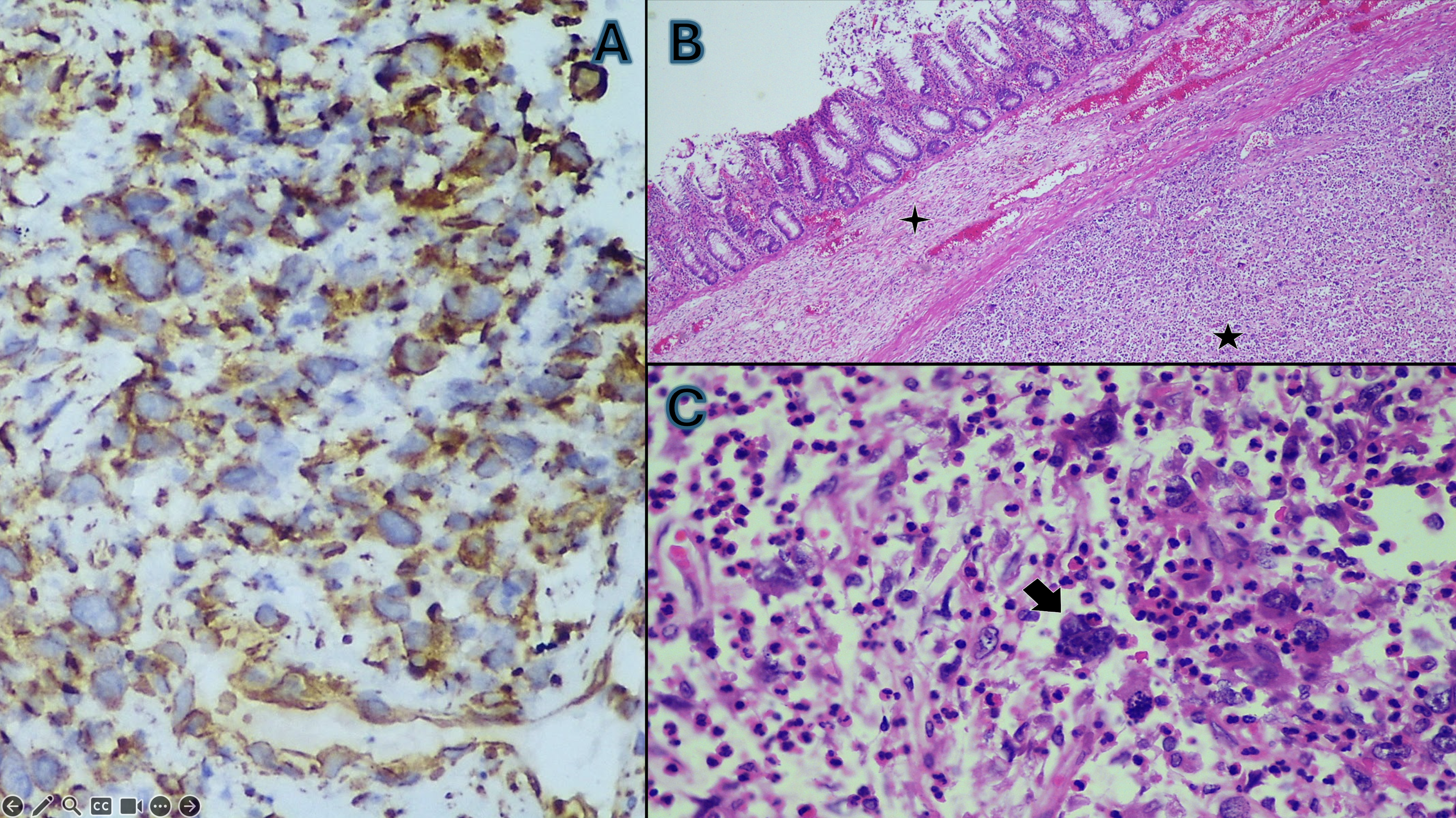 (A) Initial biopsy from colonoscopy: immunohistochemistry for vimentin showing diffuse cytoplasmic positivity in tumor cells, supporting mesenchymal lineage. (B) Resection specimen, H&E, low-power panoramic view: sarcoma (five-point star) situated beneath intact colonic mucosa (four-point star). (C) Resection specimen, H&E, high-power (40×): multinucleated tumor cell indicated by an arrow, with a prominent inflammatory infiltrate composed of eosinophils, neutrophils, and monocytes. The complete IHC panel-negative for CK AE1/AE3, CD45, CD20, CD30, CD138, CD117, DOG-1, Melan-A, ERG, smooth-muscle actin, CD21, and HHV-8, with CD3 limited to reactive lymphocytes-established UPS by exclusion.