

Danielle Krakosky, MD
Resident
University of Washington
Seattle, Washington, United States
Disclosure information not submitted.
dirir abdullahi, MD
Fellow
University of Washington
University of Washington
seattle, WA, United States
Disclosure information not submitted.
Alexa T. Andre, MD
Resident
University of Washington
Seattle, Washington, United States
Disclosure information not submitted.
.jpg "Mukta Krane, MD (she/her/hers) photo")
Mukta Krane, MD (she/her/hers)
Associate Professor of Surgery, Chief of Section of Colorectal Surgery
University of Washington
University of Washington Medicine
Seattle, WA, United States
Disclosure information not submitted.
 photo")
Sarah J. Atkinson, MD (she/her/hers)
Assistant Professor
University of Washington
University of Washington
Seattle, WA, United States
Disclosure information not submitted.
.png) Patient presented with the symptoms as seen in box 1, underwent appropriate diagnostic work up, box 2 and 3, multidisciplinary discussion box 4, IR guided biopsy box 5, surgical resection box 6 followed by path report box 7
Patient presented with the symptoms as seen in box 1, underwent appropriate diagnostic work up, box 2 and 3, multidisciplinary discussion box 4, IR guided biopsy box 5, surgical resection box 6 followed by path report box 7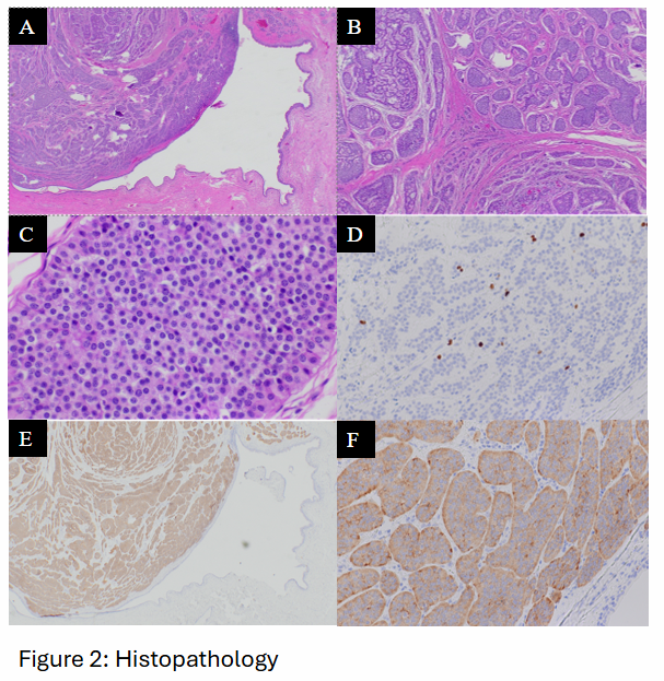 Histologic sections show monotonous neoplastic cells with coarse "salt-and-pepper" chromatin (C, 40x), minimal cytologic atypia, and a moderate amount of eosinophilic cytoplasm arranged in a nested to trabecular growth pattern (A-B, 2x-4x). Ki-67 proliferative index is ~1% (D). The neoplastic cells are diffusely positive for synaptophysin, a marker of neuroendocrine differentiation (E-F, 2x - 40x).